Figure 4.
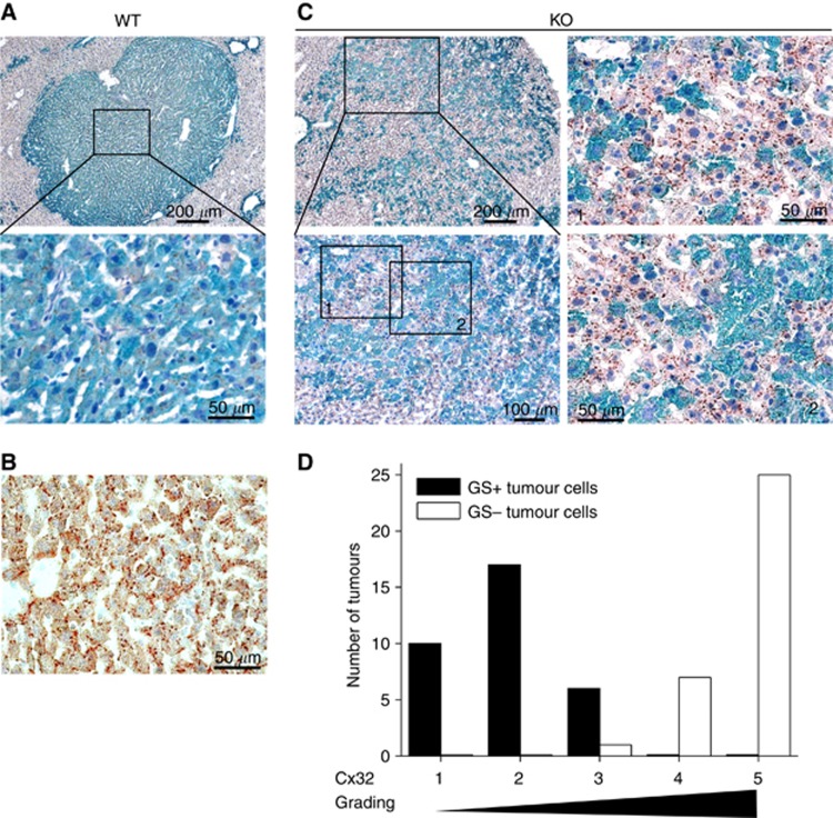
Appearance of Cx32 at membranes of Ctnnb1 KO tumour cells. (A) Reduced Cx32 levels are observable in the membranes of Ctnnb1-mutated, GS-positive tumour cells in livers from Ctnnb1 WT animals. (B) In contrast, normal liver tissue shows high membranous Cx32 levels. (C) After KO of Ctnnb1, Cx32 reappears in the GS-negative tumour cell subpopulations (image details are referred to as 1 and 2). The Ctnnb1 KO animal was killed 7 weeks after β-catenin ablation. (D) Cx32 grading of GS-positive and -negative tumour cells in hepatomas of KO mice revealed higher Cx32 levels in tumour populations lacking GS expression. Tissue slices were double stained for Cx32 and GS and analysed by light microscopy for the presence of Cx32 plaques at the cell membranes of hepatoma cells. For each tumour (in total, n=33 tumours were analysed), the degree of Cx32 expression in the two hepatoma subpopulations was classified from grade 1 (low Cx32 levels) to 5 (high Cx32 levels, comparable to surrounding normal tissue). The numbers of tumours in each class are given in the diagram for the GS-positive and GS-negative sub-populations, respectively.

