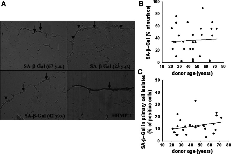
Fig. 3.
Relationship between SA-β-Gal expression in vivo and calendar age of tissue donor. Representative result of cytochemical detection of SA-β-Gal (positive cells are marked with arrows) in the omental tissue specimens; magnification ×40. The age of the tissue donors is shown in the brackets. The bottom right picture presents the results of a representative staining for the mesothelial cell-specific antigen, HBME-1 (brown area marked with an arrow) (a). Correlation between SA-β-Gal expression in the omental tissue and the calendar age of the tissue donor. The analysis was performed planimetrically and the blue SA-β-Gal-derived signal was expressed as a percentage (%) of the total mesothelium area delineated by HBME-1-derived brown staining in a corresponding section (treated as 100 %) (b). Correlation between SA-β-Gal expression in primary cell isolates from the omentum and the calendar age of the tissue donor (c). Experiments were performed on cell cultures derived from 29 different donors. (Color figure online)

