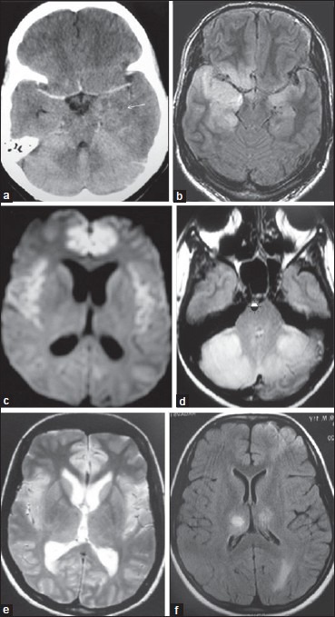
Figure 2.

(a) Computed tomography — hypo density in the left medial temporal lobe-paraneoplastic. (b) FLAIR hyper intensity both hippocampi, parahippocampi, adjacent anterior temporal lobes-Herpes simplex. (c) Autoimmune limbic encephalitis: Axial DWI reveals (d) bilateral symmetrical foci of diffusion restriction involving cingulate, medial superior frontal gyriand Insula. (e) Case of varicella encephalitis: Axial T2WI shows abnormal bilaterally symmetrical hyperintensities involving cingulate (arrow), insula and thalami. (f) Axial FLAIR imageshyperintensities involving bilateral thalami, cingulate and anterior frontal lobes
