
Figure 3.
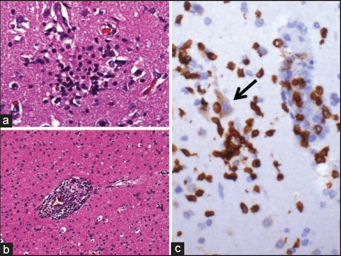
Pathology viral meningoencephalitis. (a) Microglial nodule in the grey matter marking a focus of neuronophagia. (b) White matter with perivascular inflammation. (c) CD3 immunostaining shows mainly T lymphocytes in the microglial nodule with neuronophagia (arrow)