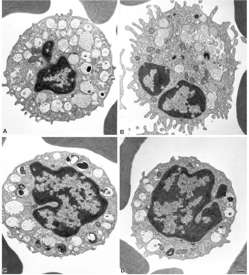
Fig. 5.
Ultrastructure of resting and activated releaser and non-releaser basophils. Releaser (A,B) and non-releaser (C,D) basophils were incubated without (A,C) and with (B,D) 1 µg/ml of anti-IgE for 30 miniutes, then washed and processed for electron microscopy as in [26]. Resting cells (A and C) show very similar membrane and granule morphologies. In releaser cells (B), FcεRI crosslinking induces granule-granule fusion, degranulation and membrane ruffling. Non-releaser cells (D) show little morphological changes following FcεRI crosslinking. Bar = 0.5 µm. Details are in [28].

