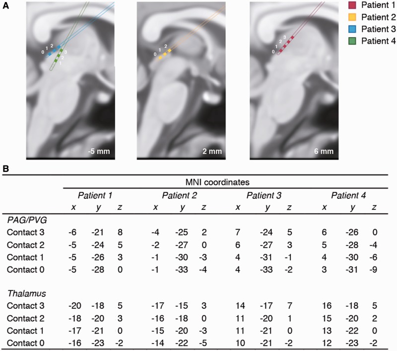
Fig. 1.
(A) Three sagittal slices of the averaged standard brain in MNI space (−5, 2 and 6 mm) showing the approximate locations of the implanted PAG/PVG electrode placements in the each of the four patients (colour coded), with each contact point numbered. Each electrode had four contact points (those points that can be shown in the present slice are in filled colour). (B) MNI co-ordinates of the electrode contact points in the PAG/PVG and sensory thalamus. For reference, typical MNI coordinates for the inferior colliculus are (x,y,z) = [6, −33, −9] and for the medial geniculate nucleus (x,y,z) = [17, −24, −2].

