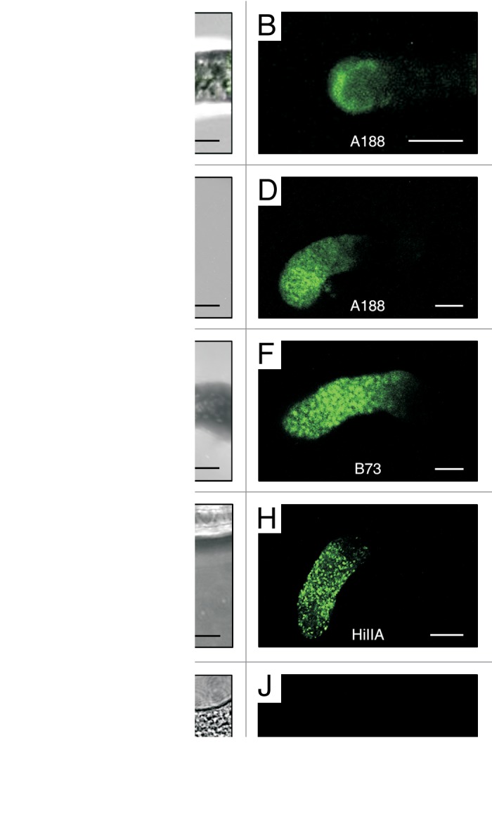
Figure 1. EA1 peptide interacts with the maize pollen tube apex in a species-specific manner. In vitro pollen tube binding assays of three maize inbred lines and the maize relative Tripsacum dactyloides (T.d.) were performed with synthetic predicted mature EA1 peptide labeled with the green fluorophore DyLight 488 NHS Ester. Z-projections of confocal image stacks of 6 to 20 μm thick sections are shown in each panel. (A, C, E, G and I) Merged bright field and fluorescence micrographs. (B, D, F, H and J) UV-fluorescence images. (A–B) Labeled EA1 fluorescence is visible at the apical membrane region of a A188 pollen tube tip. (C–H) Pollen tubes of A188, B73, and HiIIA, respectively, displaying fluorescence at their apical region in vesicle-like structures, whereas (I–J) fluorescence is not detectable from T. d. pollen tubes. Scale bars represent 10 μm.
