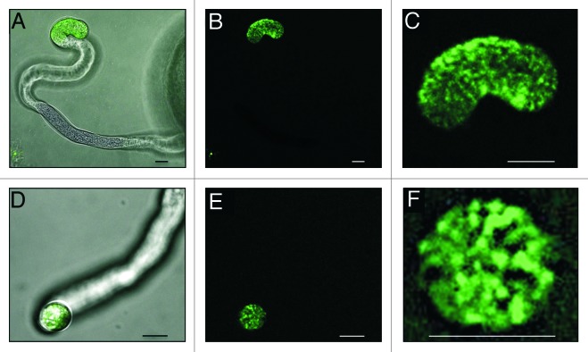
Figure 2. DyLight-labeled synthetic EA1 peptide gets internalized in vesicles at the apical region of the maize pollen tube tip. (A and D) Merged bright field and fluorescence micrographs. (B, C, E and F) UV-fluorescence micrographs. (A–B) Side-view of a pollen tube interacting with labeled EA1 peptide at its apical and sub-apical region. (C) Close-up of (B) displaying labeled EA1 in vesicle-like structures inside the pollen tube tip region. (D‒E) A pollen tube tip facing toward the observer displays labeled EA1 peptide in the tube tip. (F) Close-up of (E) displaying labeled EA1 in vesicles inside the pollen tube tip. Single optical sections are shown in (B, C, E and F). Scale bars represent 10 μm.

An official website of the United States government
Here's how you know
Official websites use .gov
A
.gov website belongs to an official
government organization in the United States.
Secure .gov websites use HTTPS
A lock (
) or https:// means you've safely
connected to the .gov website. Share sensitive
information only on official, secure websites.
