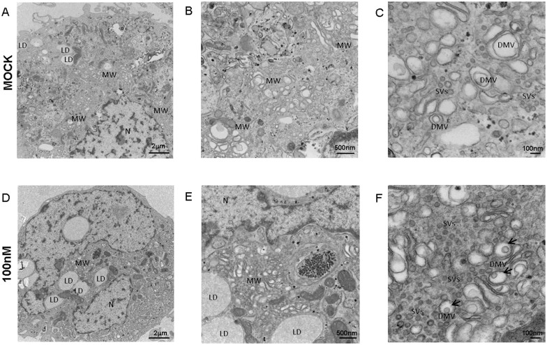
Figure 8. Electron microscopy shows that low dose treatment with inhibitor A decreases the number of membranous webs and alters their structure while having no effect on lipid droplets in Huh7-SGR cells.
Huh7-SGR cells treated for 96 h with vehicle (DMSO) (A–C) or 100 nM inhibitor A (D–F) were fixed and processed for EM. One representative cell is shown for each treatment at magnifications of 1500× (A), (D), 5000× (B), (E) and 15000× (C), (F). Treated cells generally showed more distortions in the double membrane vesicles, as shown by the black arrows. Treated cells also showed more small vesicles relative to control Huh7-SGR cells. MW: membranous webs, LD: lipid droplets, N: nucleus, DMV: double membrane vesicles, SVs: small vesicles, Arrows: reduction in inner membrane of DMVs.

