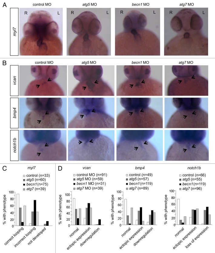
Figure 4. Autophagy gene knockdown results in abnormal cardiac looping and valve development. (A and C) In situ hybridization with a myl7 probe of morphant hearts at 48 hpf. (A) Representative images of a control morphant showing correct looping with the atrium placed left and caudal, and the ventricle right and rostral, and of autophagy morphants, showing linearized hearts without looping. (C) Quantification of data shown in (A). More than 30 embryos were analyzed in each group. (B and D) In situ hybridization of morphants with probes that detect cardiac valve markers at 2 dpf. (B) Representative images of vcan, bmp4, and notch1b expression in the hearts of control and autophagy mutants. (D) Quantification of data shown in (B). More than 30 embryos in each group were analyzed. R, right; L, left.

An official website of the United States government
Here's how you know
Official websites use .gov
A
.gov website belongs to an official
government organization in the United States.
Secure .gov websites use HTTPS
A lock (
) or https:// means you've safely
connected to the .gov website. Share sensitive
information only on official, secure websites.
