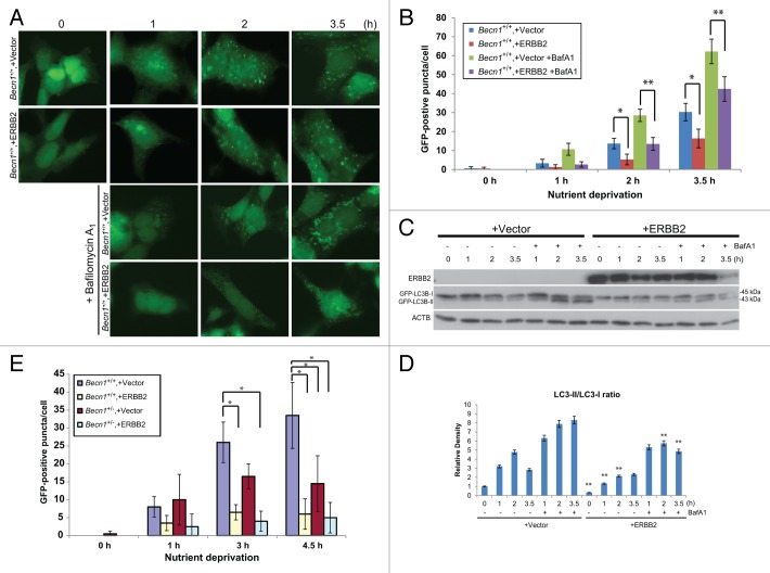
Figure 3. Transient ERBB2 overexpression inhibits stress-induced autophagy in Becn1+/+ iMMECs to the level observed in partially autophagy-defective non-ERBB2-expressing Becn1+/− iMMECs. (A) GFP-fluorescence microscopy of EGFP-LC3B-expressing Becn1+/+ iMMECs transiently transfected with a ERBB2-expressing or vector control plasmid under nutrient deprivation conditions without or with bafilomycin A1 (BafA1, 25 nM). (B) Autophagy quantification of (A) based on number of GFP-fluorescent puncta per cell. Each data point is an average of triplicate experiments ± SD after quantifying puncta in 100 cells per experiment. *P value < 0.05; **P value < 0.01. (C) GFP and ACTB western blots of whole cell protein lysates from Becn1+/+ iMMECs transiently expressing ERBB2 under nutrient deprivation without and with BafA1. (D) Densitometric analysis of LC3B-II/LC3B-I ratio, as normalized to ACTB, using ImageJ. (E) EGFP-LC3B-expressing Becn1+/+ and Becn1+/− iMMECs transiently transfected with a ERBB2-expressing or vector control plasmid were subjected to nutrient deprivation, and autophagy was quantified by the number of GFP-fluorescent puncta per cell. Each data point is an average of triplicate experiments ± SD after quantifying puncta in 100 cells per experiment. *P value < 0.05; **P value < 0.01.

An official website of the United States government
Here's how you know
Official websites use .gov
A
.gov website belongs to an official
government organization in the United States.
Secure .gov websites use HTTPS
A lock (
) or https:// means you've safely
connected to the .gov website. Share sensitive
information only on official, secure websites.
