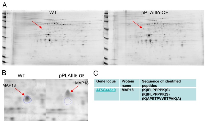
Figure 1. Comparative analysis of membrane proteins isolated from WT and OE Arabidopsis seedlings. (A) 2-D gel images of membrane proteins from one-week-old liquid-grown Arabidopsis seedlings. The plasma membrane fraction was isolated by 2-phase partitioning with 6% polyethylene glycol 3350, 6% Dextran T-500, and 8 mM KCl. The lower conductivity plasma membrane protein was prepared using the kit of perfect-FOCUS (G-BIOSCIENCE). Isoelectric focusing electrophoresis (IEF) was performed using a Bio-Rad Protean IEF cell, followed by SDS-PAGE on Bio-Rad Criterion pre-cast gels (8–16%). Gel images were acquired using a Typhoon 9410 variable mode imager (Amersham Biosciences, Pittsburgh, PA). Each gel was imaged sequentially at excitation/emission filter wavelengths. Image analysis was performed using SameSpots software (Nonlinear Dynamics, Durham, NC) for gel alignment, spot averaging and normalization. (B) One representative of 3 gel images indicating the mobility shift of a protein spot that subsequently identified as MAP18. Protein spots of interest were proteolytically digested and sequenced using an ABI QSTAR XL (Applied Biosystems/MDS Sciex) hybrid QTOF MS/MS mass spectrometer equipped with a nanoelectrospray source.

An official website of the United States government
Here's how you know
Official websites use .gov
A
.gov website belongs to an official
government organization in the United States.
Secure .gov websites use HTTPS
A lock (
) or https:// means you've safely
connected to the .gov website. Share sensitive
information only on official, secure websites.
