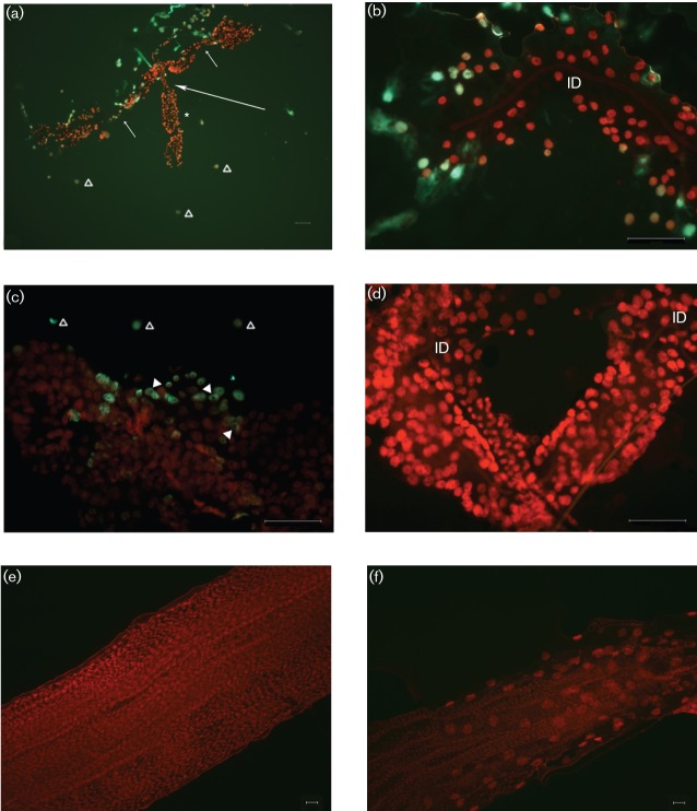
Fig. 1.
Epifluorescent micrographs of whole-mount preparations of SGs, midgut and hindgut from adult female A. albopictus mosquitoes, following injection with high-titre SINV (25 500 p.f.u. per insect). Tissues were stained using the TUNEL assay to detect apoptotic cells (green) and counterstained with propidium iodide (red). (a) Apoptosis was associated with the proximal and distal regions of two lateral lobes at day 5 p.i. (short arrows; neck region separating proximal and distal lateral lobes). Apoptosis was not detected in median lobes (*, long arrow; neck region of median lobe). Gross pathology (distension and cellular disruption) was observed in lateral lobes but not in the median lobe. Apoptotic-positive cells scattered around the SGs (▵). (b) Distal region of lateral lobe at day 5 p.i. Note apoptotic-positive cells integral to the lobe epithelium. Note the overall low cell density compared with the uninfected lobe (d) and absence of apoptosis in the internal duct (ID). (c) Distal region of lateral lobe at day 7 p.i. Note apoptotic-positive cells integral to lobe (arrowheads; indicate cytoplasmic staining) and apoptotic-positive cells scattered around SGs (▵). (d) Distal region of lateral lobe from an uninfected mosquito at day 5 p.i. Note overall high cell density compared with infected tissue (b, c). Apoptosis was not detected in the SG parenchyma, internal ducts (ID) or in cells scattered around the SGs (compare with a and c). Midgut (e) and hindgut (f) from infected mosquitoes (day 5 p.i.). Note the absence of apoptosis (green) in both gut regions. Bars, 50 µm (a–d); 10 µm (e, f).

