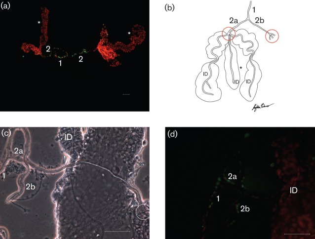
Fig. 2.
Whole-mount SG preparations from uninfected A. albopictus mosquitoes. SG common duct (1) and main ducts (2; 2a, 2b). Triad structure (circled) indicates the junction where the main ducts (2) branch into the internal ducts (ID). (a) Epifluorescent image stained with the TUNEL assay; SGs with ducts intact. Note the presence of apoptosis (green) associated with the common duct (1) and main ducts (2). *, Median lobes. (b) Graphic drawing for orientation. (c) Phase-contrast image for orientation. (d) Apoptotic cells (green) outline the common (1) and main (2a, 2b) ducts, but are not observed in the internal ducts (ID). Bars, 10 µm (a); 50 µm (c, d).

