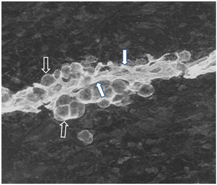
Figure 3.
Immunofluorescence of subcutaneous adipose tissue from a 75 day old pig fetus. Cryostat sections were stained for the AD-3 monoclonal antibody (Mab) and then incubated with FITC-conjugated goat anti-mouse IgG3. Stained sections were mounted in Elvanol and examined using a fluorescence microscope. Immunoreactive capillaries and adipocytes are obvious in a developing adipocyte cluster after the onset of adipocyte differentiation in the fetus. Immunoreactivity for AD-3, a preadipocyte marker, clearly indicates that both of the adipocytes (black arrow, white trim), associated capillaries (white arrow, black trim) and probably perivascular cells express the AD-3 antigen indicating a common lineage for capillary endothelial cells preadipocytes and perivascular cells. Note the close proximity of adipocytes and capillaries indicating that the proximity was even greater earlier in development. Note the absence of immunoreactivity in areas distal to the adipocyte cluster.Staining was not observed using the FITC-conjugated second antibody alone or in conjugation with an isotype-matched control MAb.

