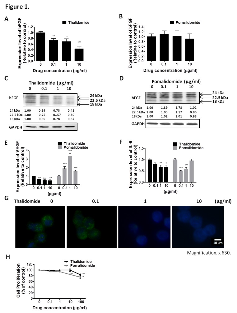
Figure 1. Effect of thalidomide or pomalidomide on bFGF, VEGF and IL-6 expression in RPMI8226 cells.

RPMI8226 cells were treated with (A) thalidomide or (B) pomalidomide for 4 hours. The bFGF mRNA expression was monitored by real-time PCR, with the expression of GAPDH used as an internal control. Protein extracts from RPMI8226 cells treated with (C) thalidomide or (D) pomalidomide for 4 hours were subjected to Western blot for analysis of bFGF protein level. GAPDH is the internal control. RPMI8226 cells were treated with thalidomide and pomalidomide for 4 hours and mRNA levels of (E) VEGF and (F) IL-6 were monitored by real-time PCR. (G) Thalidomide regulates bFGF expression and cellular distribution in RPMI8226 cells. Immunofluorescence detection of bFGF in RPMI8226 cells treated with thalidomide for 4 h. Cellular distribution of bFGF was studied by fluorescence microscopy. DNA was stained with H33258 as a nuclear marker. Magnification, × 630. (H) Viable cell counts using trypan blue. Cell viability of RPMI8226 cells treated with thalidomide and pomalidomide. RPMI8226 cells, which were treated with the indicated concentrations (0, 0.1, 1 and 10 μg/mL) of thalidomide and pomalidomide for 72 hours, were stained with trypan blue. Data were collected from at least 3 independent experiments. The results were expressed as the relative index of untreated control ± S.E.M. (*, P < 0.05, **, P < 0.01, ***, P < 0.001, Student's t-test).
