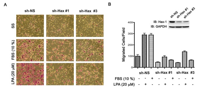
Figure 2. Silencing of Hax-1 attenuates LPA and FBS stimulated invasive migration of SKOV3 cells.
(A) Transient silencing of Hax-1 was carried out by transfecting SKOV3 cells with vectors encoding shRNA specific for Hax-1 (sh-Hax 1 & sh-Hax #3) or scrambled shRNA control for 48 hours (sh-NS). These transfectants were unstimulated (SS), stimulated with LPA (20 μM), or FBS (10%) and their migratory responses were monitored as described under Materials and Methods. At 24 hours following stimulation, images were obtained from random fields of view at 10X magnification. The images shown are representative of three independent experiments, each performed with triplicate fields of view. (B) Cell migration profiles were quantified by enumerating the migrated cells in a minimum of three different fields. Results are presented as the number of migrated cells per field and the bars represent mean ± SEM from there independent experiments. Silencing of endogenous Hax-1 was monitored by immunoblot analysis using antibodies to Hax-1 (inset). The blot was stripped and reprobed with antibodies to GAPDH to monitor equal loading of protein.

