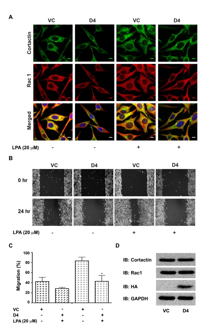
Figure 7. Expression of Hax-D4 attenuates LPA stimulated Rac1-cortactin complex formation in SKOV3 Cells.
(A) pcDNA3-SKOV3 vector control cells (VC) and pcDNA3-Hax D4 domain transfected cells (D4) were serum starved for 16 hours and stimulated with 20 μM LPA for 24 hours. At the end of 24 hours, the cells were immunostained for Rac 1 and cortactin following previously discussed procedures. Fluorescent micrographs were collected with a Leica SP2 MP Confocal microscope using 63x Plan APO 1.4 NA oil immersion objective. Colocalization of Rac and Cortactin (Yellow) were analyzed, on 3 images for each condition per experiment, using NIH ImageJ software. The scale bar 10 μm is common for all images. (B) SKOV3 cells were transiently transfected with pcDNA3-SKOV3 vector control (VC) and pcDNA3-Hax D4 domain. 24 hours after transfection, transfectants were serum starved for 16 hours and linear scratch wounds were made across the monolayers to assay the wound healing efficiency of the cells. Random fields of view (10X) were photographed, marked for re-identification and stimulated with 20 μM LPA and identical fields were re-imaged following 24 hours. The experiments were performed thrice independently and presented images are from a typical experiment. (C) The percentage of wound closure was calculated based on the migration of the control and the D4 domain transfected cells (mean ± SEM; n = 3). The statistical significance was assessed using one-tailed t-test. * p<0.05. (D) 50 μg of lysate protein from D4 domain transfected cells was resolved using 15% SDS-PAGE and immunoblot analysis was carried out with antibodies against Rac1, cortactin and HA-epitope. The blot was stripped and reprobed with an antibody to GAPDH for ensuring equal protein loading.

