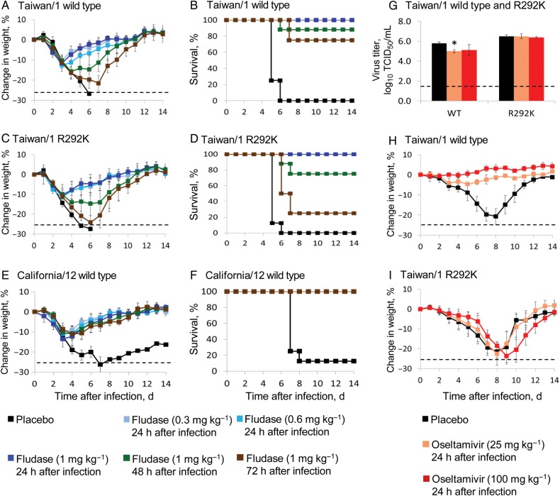
Figure 2.
Efficacy of delayed treatment in mice. Animals (8/group) were intranasally inoculated with 5 50% mouse lethal doses of plaque-purified influenza A virus subtype H7N9, equivalent to 1.6 × 10⁵ 50% tissue culture infective doses (TCID50) of Taiwan/1 wild-type (A, B) or 1.6 × 10⁵ TCID50 of Taiwan/1 R292K (C, D). California/12 (a 2009 pandemic influenza A virus subtype H1N1 isolate; 5 × 10⁴ TCID50) was used as a control (E, F). Mice were treated with DAS181 24, 48, or 72 hours after infection (for a total of 6 regimens). In oseltamivir control groups, mice (n = 8) were infected with 104 TCID50 of Taiwan/1 wild-type (G, H) or R292K (G, I) virus and received twice-daily treatment for 5 days, starting 24 hours after infection. Three mice were euthanized on day 6 after infection, and virus titers in lungs were determined by the TCID50 assay in MDCK cells (G); dotted lines indicate the lower limit of virus titer detection (1.6 log10 TCID50/mL). Body weights were monitored daily (A, C, E, H, I); animals that lost ≥25% (shown in dotted lines) of their initial body weights were humanly euthanized, and numbers of animals that survived are shown (B, D, F). P values of <.05 (asterisks) denote statistically significant differences from values for placebo-treated groups. Error bars indicate SDs.

