
Figure 2.
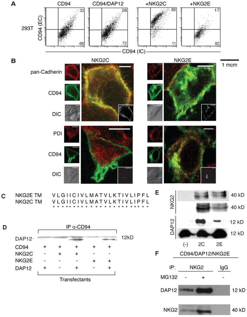
NKG2E forms an intracellular complex with DAP12 and CD94, and is trafficked to the ER but not the plasma membrane. (A) 293T cells were transfected with CD94 alone (first panel), CD94 and DAP12 (second panel), CD94/DAP12/NKG2C (third panel), or CD94/DAP12/NKG2E (fourth panel). Cells were stained for surface expression of CD94 and were permeabilized and stained for intracellular CD94. Data are representative of at least three independent experiments. (B) 293T cells were transfected with CD94, DAP12, and either NKG2C (left panels) or NKG2E (right panels) and assessed by confocal microscopy. CD94 is shown in green while differential interference contrast (DIC) is shown in gray for all samples. Pan-cadherin is shown in red in the top two panels, while protein disulfide-isomerase (PDI) is shown in red on the bottom two panels. Images are representative of at least three independent experiments. (C) NKG2C and NKG2E have identical transmembrane domains, suggesting that NKG2E can associate with CD94. (D) DAP12 associates with CD94 in the presence of NKG2C or NKG2E in 293T cells. 293T cells were transfected as in (B). Co-immunoprecipitation was performed on lysates, using an anti-CD94 mAb to pull down the NKG2/CD94/DAP12 complex, which was then probed using an anti-DAP12 mAb. Data are representative of at least three independent experiments. (E) NKG2E forms a complex with DAP12 and CD94 in Ba/F3 cells. Ba/F3 cells were transfected as in (B). Cell lysates were immunopreciptated with anti-CD94 and probed with anti-NKG2 (top row). Lysates were also immunoprecipitated with anti-NKG2 and probed with anti-DAP12 (third row). Total lysates were blotted with anti-NKG2 (second row) and anti-DAP12 (bottom row) as loading controls. Data are representative of at least three independent experiments. (F) Proteasomal inhibitor MG132 increases expression of CD94/NKG2E/DAP12 complexes. Ba/F3 cells were transfected with CD94, DAP12, and NKG2E, and were then treated with MG132 or with a vehicle control. Lysates were subsequently immunoprecipitated using an anti-NKG2 mAb or an IgG control. The resulting complex was probed using an anti-DAP12 mAb (top row) or an anti-NKG2 mAb (bottom row). Data are representative of at least three independent experiments.