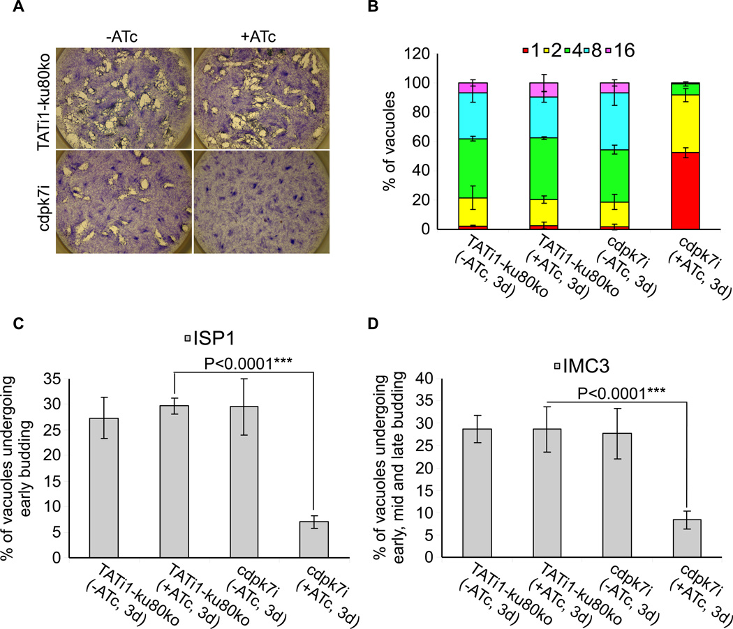
Figure 3. Phenotypic consequences of TgCDPK7 depletion in cdpk7i strain.
(A) Plaque assay performed on HFF monolayer infected with TATi1-ku80ko or cdpk7i parasites pretreated first during 48 hours with ATc. After 7 days ± ATc, the HFF were stained with Giemsa. (B) Intracellular growth of TATi1-ku80ko and cdpk7i cultivated in presence or absence of ATc for 48 hours and allowed to invade new HFF cells. Numbers of parasites per vacuole (X axis) were counted 24 hours after inoculation. The percentages of vacuoles containing varying numbers of parasites are represented on the Y-axis. Values are means ± SD for three independent experiments. (C and D) Endodyogeny assay performed on TATi-ku80ko or cdpk7i strains cultivated in presence or absence of ATc for 48 hours and allowed to invade new HFF cells. Numbers of vacuoles showing the formation of newly formed buds (Y axis) were counted 24 hours after inoculation using anti-ISP1 or anti-IMC3 antibodies. Values are means ± SD for three independent experiments. Statistical significance was evaluated using the student’s t test. ***P<0.0001 (C, ISP1), ***P<0.0001 (D, IMC3).

