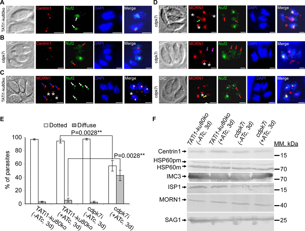
Figure 7. TgCDPK7 knockdown affects the distribution of MORN1 and Nuf2 proteins.
(A) to (D): IFA analysis of parasites undergoing division after 3 days of ATc treatment. (A) and (C): IFAs of representative TATi1-ku80ko parasites which were compared to IFAs of representative cdpk7i mutant parasites, (B) and (D). (A) and (B): anti-centrin1 (in red) and anti-Nuf2 (in green) antibodies were used to stain the centrosome and kinetochore respectively. (A) Two TATi1-ku80ko vacuoles were shown with each containing 1 parasite undergoing division. The kinetochore (in green) in each parasite is marked with a white arrow. TgNuf2 protein which is a component of the kinetochore complex is concentrated in the parasite nucleus and is flanked by two centrosomes during division (white arrows). (B) A cdpk7i vacuole showing the typical dotted-like staining of Nuf2 protein surrounded by a duplicated centrosome in only 1 parasite out of 4. In the parasites displaying a stretched centrosome or an undetectable centrin 1 labelling, the Nuf2 protein was found diffused throughout the nucleus (pink arrows). (C) and (D): anti-Myc (TATi1-ku80ko and cdpk7i parasites express a Myc tagged MORN1 protein) and anti-Nuf2 antibodies were used to label the spindle pole and kinetochore sub-cellular structures respectively. (C) A TATi1-ku80ko vacuole containing 4 parasites is shown. The kinetochore (in green) in each parasite is marked with a white arrow. TgNuf2 protein (white arrows) shows a partial colocalisation with MORN1 protein which is a component of the spindle pole structure located at the nuclear envelope of the parasite (blue arrows). The MORN1 protein marked also the basal end of the parasites (white asterisks). (D) IFA showed abnormal MORN1 protein distribution at the spindle pole in cdpk7i parasites displaying a diffused Nuf2 protein (pink arrows). The MORN1 protein was distributed in the form of a pipe (green arrow), was found undetectable (the two white arrows) or diffused (yellow arrow). Scale bars represent 2 µm. (E) Scoring of Nuf2 protein distribution defect by IFA using anti-Nuf2 antibodies. Data are mean values ± SD for three independent experiments. Statistical significance was evaluated using the student’s t test. **P=0.0028 (dotted), **P=0.0028 (diffuse). (F) Western blot analyses performed on mutant or TATi1-ku80ko parasite lysates probed with anti-centrin1 or anti-HSP60 or anti-IMC3 or anti-ISP1 or anti-Myc or anti-SAG1 antibodies. pm: pre-mature; m: mature.

