Figure 2.
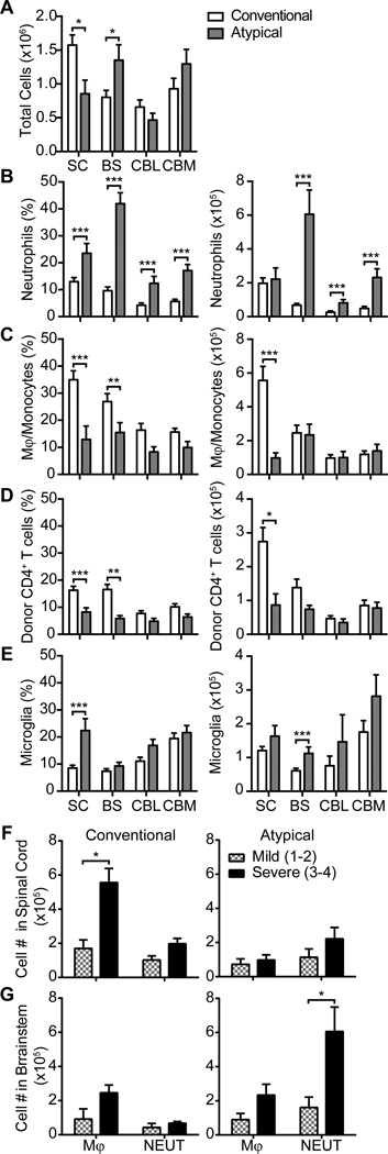
Neutrophils are prominent in the brainstem of IFNγRO mice with atypical EAE while monocytes and donor T cells are prominent in the spinal cord of WT mice with conventional EAE. (A) The average number of total cells isolated from the spinal cord (SC), brainstem (BS), cerebellum (CBL), or cerebrum (CBM) of WT mice with pure conventional and IFNγRO mice with pure atypical EAE. All animals had moderate to severe disease (clinical scores of 3–4) at the time of euthanasia. (B–E) Flow cytometry was performed to enumerate the percent and number of infiltrating neutrophils (CD11b+CD45+Ly6G+) (B), monocytes/ macrophages (CD11b+CD45hiLy6G−) (C), donor CD4+ T cells (CD3+CD4+CD45.1+)(D), and microglia (CD11b+CD45intLy6G−) (E).
(F and G) The absolute number of monocytes (MONO) and neutrophils (NEUT) per spinal cord (F and brainstem (G) were compared between mice with mild (clinical scores 1–2) or severe (clinical scores 3–4) EAE. Data were pooled from at least 3 experiments with a total of 27 WT and 20 IFNγRKO mice. Flow cytometry gating scheme is illustrated in Fig. S2. *P<.05, **P<.01,***P<.001

