Figure 1.

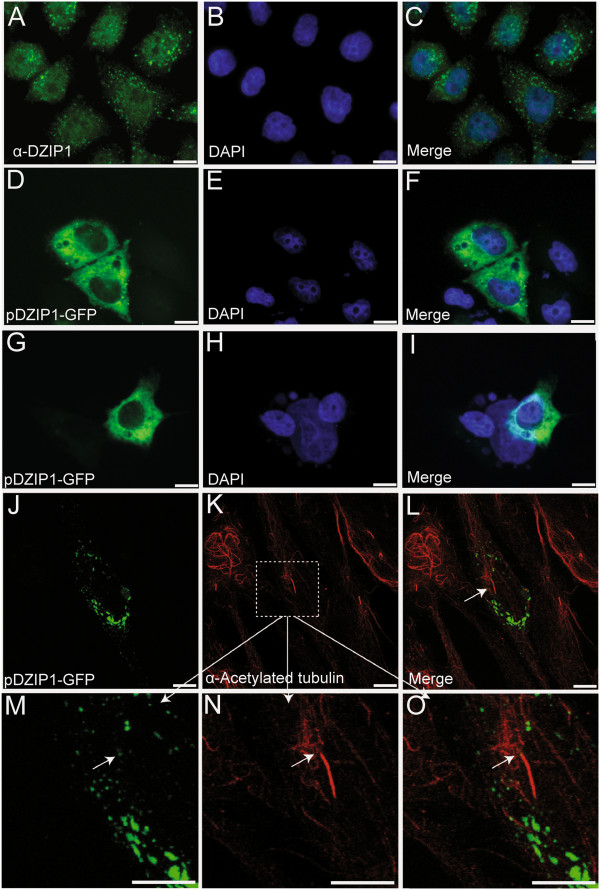
DZIP1 is located predominantly in the cytoplasm and shows a granular distribution. (A) Indirect immunofluorescence staining of DZIP1 (green) in HeLa cells. (B) Nuclei counterstained with DAPI (blue) and (C) merged image. HeLa (D-F), HEK293 (G-I) and hTERT-RPE1 (J-O) cells were transfected with pDZIP1-GFP. (J-O) Ciliary axonemes were labeled with anti-acetylated tubulin antibodies (red). (M-O) are magnified sections from (K) (white boxes). Arrow = potentially compatible with a basal body localization. Scale bar: 10 μm.
