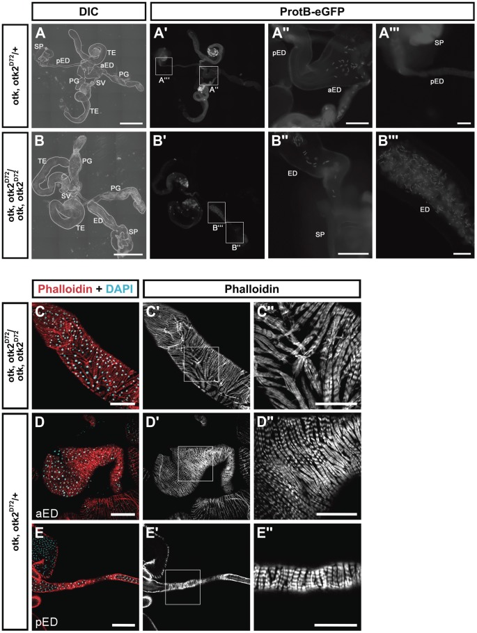
Figure 7. otk, otk2 loss of function causes malformation and obstruction of the ejaculatory duct.
(A) Overview of the reproductive tract of a male heterozygous for otk, otk2D72 and carrying a Protamin B-eGFP transgene. (A″) and (A‴) show higher magnifications of the insets highlighted in (A′). (B) Overview of the reproductive tract of a male homozygous for otk, otk2D72 and carrying a Protamin B-eGFP transgene. Note that the ejaculatory duct is severely shortened and thickened compared to (A). Also note that sperm marked by Protamine B-eGFP (B′) accumulates in the ejaculatory duct of the homozygous mutant male. (B″) and (B‴) show higher magnifications of the insets highlighted in (B′). (C) Disorganization of the muscle sheath of the ejaculatory duct in otk, otk2 homozygous mutant males. (C″) Enlarged view of the boxed area in (C′). (D, E) The muscle sheath of the anterior (D) and posterior (E) ejaculatory duct from heterozygous control males. (D″, E″) Enlarged views of the boxed areas in (D′, E′). Fluorescent Phalloidin was used to stain F-actin. aED, anterior ejaculatory duct; pED, posterior ejaculatory duct; PG, paragonium (accessory gland); SP, sperm pump; SV, seminal vesicle; TE, testis. Scale bars: A, B = 500 µm, A″, B″ = 100 µm, A‴, B‴ = 50 µm, C–E = 100 µm, C″–E″ = 50 µm.

