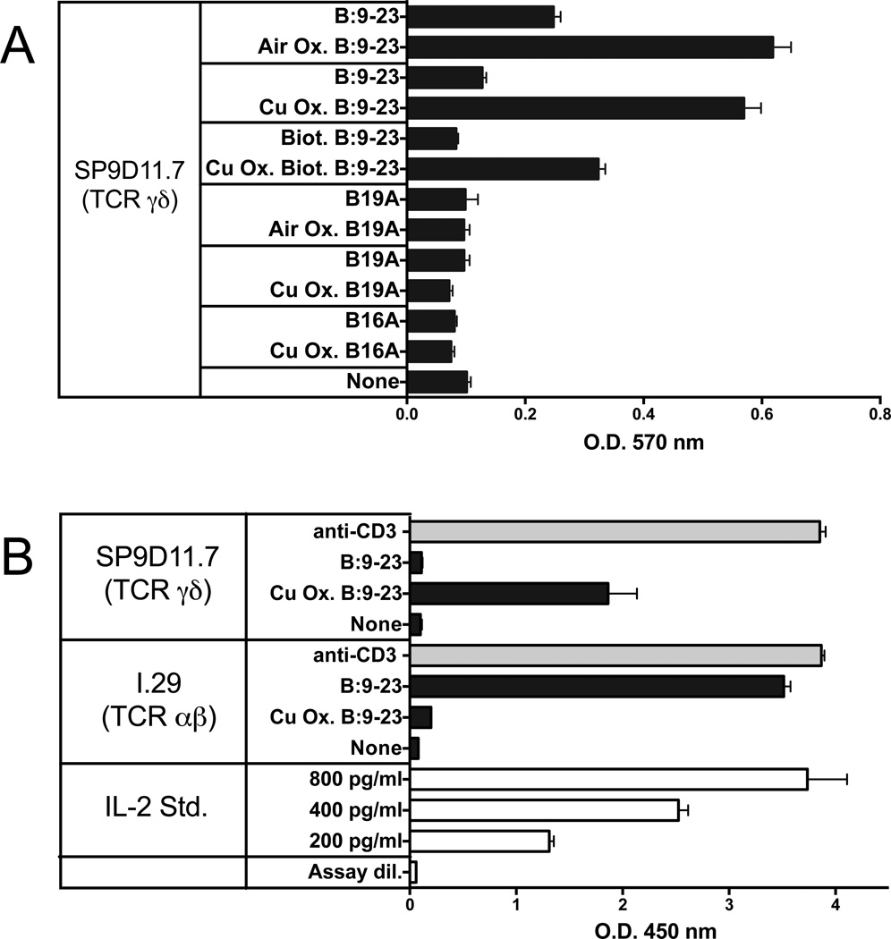
Figure 1. Oxidation of the B:9-23 peptide antigen enhances stimulation of antigen-specific γδ T cell hybridomas while reducing stimulation of an antigen-specific αβ T cell hybridoma.
Panel A Responses of hybridoma SP9D11 to untreated and oxidized peptide antigens
3×10⁴ hybridoma cells were cultured overnight either alone (none) or with untreated or oxidized (Air Ox., oxidized by exposure to ambient air, Cu Ox., oxidized with copper (II) chloride) peptide antigens at 100 µg/ml. Cellular responses were measured in triplicate using the LacZ stimulation assay. Bars show mean response values +/− SE.
Panel B Comparison of the responses of the γδ hybridoma SP9D11 and the αβ T cell hybridoma I.29 to untreated and oxidized B:9-23 peptide antigens
Culture conditions and peptide stimulation were as described for panel A, except for the addition to all cultures of 1×10⁵ fixed APCs. Cellular responsiveness was determined by stimulation with plate-bound anti-CD3ε mAbs. Cellular responses were measured in triplicate using ELISA for IL-2. Bars show mean response values +/− SE.

