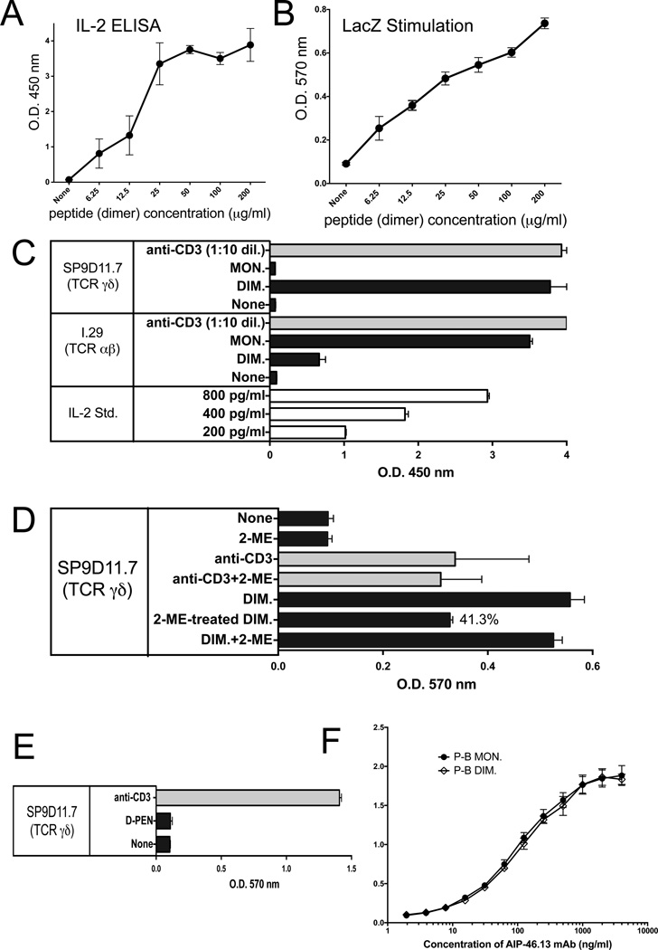
Figure 2. The γδ T cell hybridoma SP9D11 specifically recognizes oxidized dimers of the B:9-23 peptide antigen whereas the αβ T cell hybridoma I.29 recognizes monomers.
Panels A, B Response of SP9D11 to titrated amounts of dimeric B:9-23 peptide measured via IL-2 ELISA and LacZ assays, respectively.
Culture conditions and response measurements were as described in Fig.1.
Panel C SP9D11 γδ T cells selectively respond to dimers of the B:9-23-peptide, whereas I.29 αβ T cells respond to monomers.
Culture conditions, peptide stimulation and response measurements as described in Fig.1, panel B, except that HPLC-purified monomeric B:9-23 peptide (MON.) and DMSO-oxidized dimeric B:9-23 peptide (DIM.) were used as peptide antigens. Cellular responsiveness was determined by stimulation with plate-bound anti CD3ε mAbs. Supernatants of cultures with anti CD3ε were diluted 10× prior to response measurements. Bars show mean response values +/− SE.
Panel D SP9D11 Treatment of the dimeric B:9-23-peptide with 2-ME reduces its stimulatory activity.
Culture conditions, peptide stimulation and response measurements as described in Fig.2, panel C. None: no stimulation. 2-ME: 2-ME added to the cultures to a final concentration of 0.25 mM. anti-CD3: soluble anti CD3ε mAb added. Anti-CD3+2-ME: soluble anti CD3ε mAb added as well as 0.25 mM 2-ME. DIM: dimeric B:9-23 peptide added at 100 µg/ml. 2-ME-treated DIM: dimeric B:9-23 peptide pretreated with 5mM 2-ME, then added to stimulation cultures to a final peptide concentration of 100 µg/ml and a final 2-ME concentration of 0.25 mM. DIM+2-ME: dimeric B:9-23 peptide added at 100 µg/ml and 2-ME added at 0.25 mM. Cellular responses were measured in triplicate using the LacZ stimulation assay. Bars show mean response values +/− SE.
Panel E SP9D11 γδ T cells fail to respond to the penicillamin adduct of B:9-23
Culture conditions, stimulation and response measurements were as described in Fig.1, panel A. The B:9-23-penicillamin adduct was added at 100 µg/ml. Cellular responsiveness was determined by stimulation with plate-bound anti-CD3ε mAbs. Responses were measured in triplicate using the LacZ stimulation assay. Bars show mean response values +/− SE.
Panel F Monoclonal antibody AIP-46.13 recognizes monomeric and dimeric B:9-23 peptide (binding assay)
High protein binding ELISA plates were coated with purified monomeric or dimeric B:9-23 peptide at 3 µg peptide/ml. Titrated amounts of AIP-46.13 mAb were added and plate-bound antibody detected by ELISA. Curves show mean absorbance values of triplicate determinations +/− SE.

