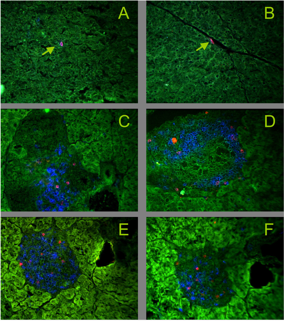
Figure 8. Vγ1+ γδ T cells infiltrate the islets of Langerhans in NOD mice.
Snap frozen pancreas tissue was acetone dehydrated and stained with mAbs. Green: tissue autofluorescence.
Panels A, B Control: C57BL/6, blue: CD3ε, red TCR-δ, arrows: individual γδ T cells, not in the islets
Panels C–F NOD, C: 16 wks of age, blue: CD3ε, red: TCR-δ; D: 8 wks of age, blue: CD3ε, red: TCR-Vγ1; E: 12 wks of age, blue: CD3ε, red: TCR-Vγ1; F: 12 wks of age, blue: CD8α, red: TCR-Vγ1

