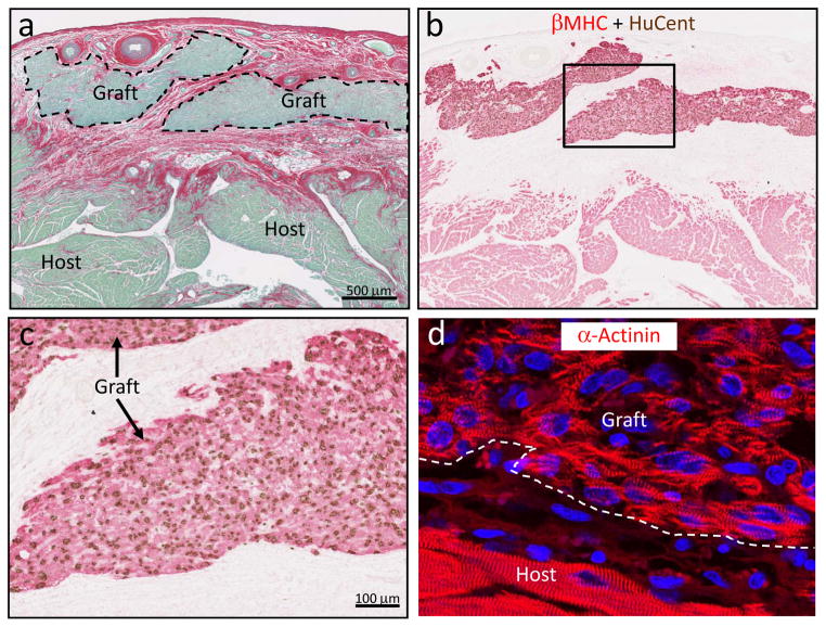
Figure 3. Grafts of human ESC-derived cardiomyocytes in the cryoinjured guinea pig heart.
Representative photomicrographs demonstrating substantial implants of human myocardium within the scar tissue. a, By picrosirius red stain, the scar appears red and viable tissue green. Scale bar=500 μm. b, The human origin of the graft myocardium was confirmed in an adjacent section by combined in situ hybridization with a human-specific pan-centromeric (HuCent, brown) probe and β-myosin heavy chain (βMHC, red) immunohistochemistry. c, Inset from panel b at higher magnification. Note the nuclear localization of the HuCent signal, confirming human origin of these cells. d, Immunostaining for α-actinin highlights the sarcomeric organization of the graft myocytes.

