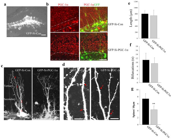
Figure 4. PGC-1α is required for the maintenance of dendritic spines in adult mouse hippocampal dentate gyrus granule neurons.
(a) Low magnification images showing GFP fluorescence in neurons in the dentate gyrus. Adenoviruses that contain GFP-Si-Con or GFP-Si-PGC1α were injected into the dentate gyrus of the hippocampus of 2 month-old male mice. Two weeks later the mice were euthanized, and brains were sectioned and confocal images of GFP fluorescence were acquired. (b) Representative confocal images showing PGC-1α immunostaining (red) and GFP (green) in the dentate gyrus of the hippocampus of mice in which granule neurons were infected with either Ad-GFP-Si-Con or Ad-GFP-Si-PGC1α. (c, d) Representative confocal images showing dendritic trees (c) and dendritic spines (d) of neurons infected with either GFP-Si-Con or GFP-Si-PGC1α. The dendritic spines were quantified in the secondary and tertiary segment of dendrites which are illustrated in b (bracelets). Scale bars in (a)=100 μm, (b) = 50 μm and in (c, d) =10 μm. (e–g) Results of quantitative analysis of total dendritic length, number of bifurcations, and dendritic spine density in dentate granule neurons infected with either Ad-GFP-Si-Con or Ad-GFP-Si-PGC1α. Values are the mean ± SD (n = 5 mice; 15–20 neurons analyzed per condition. **p<0.01 (Student’s t-test).

