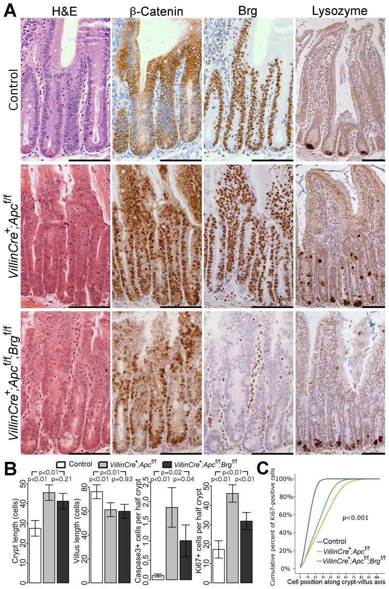
Figure 1. Brg1 loss attenuates the effects of Apc deletion in the small intestinal epithelium.
A. H&E, β-catenin and Brg1 staining of the small intestinal epithelium from control VillinCreERT2−, VillinCreERT2+Apcfl/fl and VillinCreERT2+Apcfl/flBrgfl/fl mice 4 days after high-dose induction revealed disturbed crypt architecture and nuclear localisation of β-catenin in both cohorts, as well as complete loss of Brg1 in VillinCreERT2+Apcfl/flBrgfl/fl epithelium. Lysozyme immunostaining showed mis-localisation of Paneth cells in VillinCreERT2+Apcfl/fl epithelium, which was restored to normal by inactivation of Brg1. Scale bars represent 100 µm. B. Scoring of the crypt and villus length revealed no differences between double knock-out mice (black) and VillinCreERT2+Apcfl/fl animals (grey). Scoring of the cleaved Caspase3 positive cells and Ki67 positive cells detected a decrease in both apoptosis and proliferation in VillinCreERT2+Apcfl/flBrgfl/fl mice compared to VillinCreERT2+Apcfl/fl animals. Data are shown as mean ± group's standard deviation for Caspase3 quantification and as mean ± pooled standard deviation otherwise, p value was calculated by means of t test not assuming equal variance for Caspase3 data and by means of one way ANOVA otherwise, p value was adjusted for multiple testing. For all comparisons n≥4. C. Analysis of cumulative frequency of Ki67 positive cells at each position along crypt-villus axis revealed expansion of the proliferative compartment in both double knock-out (green line) and VillinCreERT2+Apcfl/fl (orange line) mice compared to VillinCreERT2− controls (blue line). This expansion was less pronounced in double knock-out mice compared to VillinCreERT2+Apcfl/fl animals. For all pair wise comparisons Kolmogorov-Smirnov test p<0.001, n = 4.

