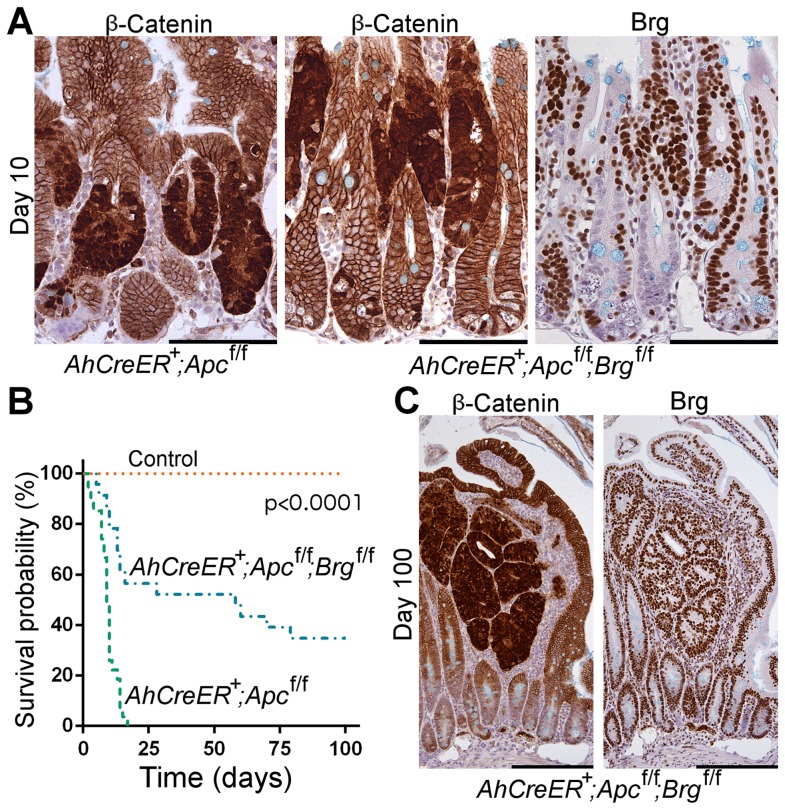
Figure 3. Brg1 loss is incompatible with Wnt-driven adenoma formation and improves animal survival upon Apc inactivation in the small intestine.
A. Immunohistochemical analysis of β-catenin revealed numerous lesions with nuclear localisation of β-catenin in the small intestine of AhCreERT+Apcfl/fl and AhCreERT+Apcfl/flBrgfl/fl mice 10 days post induction. Brg1 immunostaining of serial sections showed clusters of Brg1-deficient cells in AhCreERT+Apcfl/flBrgfl/fl intestine, but failed to detect any overlap between lesions with nuclear β-catenin and Brg1 negative cells. B. Survival analysis demonstrated significantly increased survival probability of AhCreERT+Apcfl/flBrgfl/fl mice (blue line, median survival 58 days) compared to AhCreERT+Apcfl/fl animals (green line, median survival 9 days). Log-rank test p<0.0001, n≥20. No control animals died within the timeframe of the experiment (n = 8). C. β-catenin immunostaining of the small intestinal epithelium of AhCreERT+Apcfl/flBrgfl/fl mice 100 days post induction revealed numerous micro adenomas enclosed within distended villi. Staining with Brg1 antibody showed that all the micro adenomas retained Brg1 expression. Scale bars represent 100 µm (A) and 200 µm (B).

