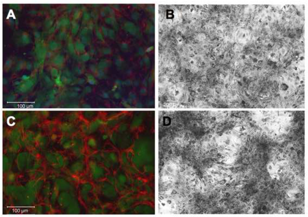
Figure 1. Heparin treatment of primary hAoSMC shows organized elastin fibers.
Adult human aortic SMCs were grown in the absence (A,B) or presence (C,D) of 300 µg/ml heparin for 7 days, then fixed and processed for immunofluorescence microscopy. Non-permeabilized samples were labeled with polyclonal anti-elastin antibody (red), CellTracker Green for cell membrane, and DAPI (blue) for nuclear stain (A,C); or with van Gieson stain for elastin (B,D). Significant extracellular elastin fibers are shown in the heparin treated samples.

