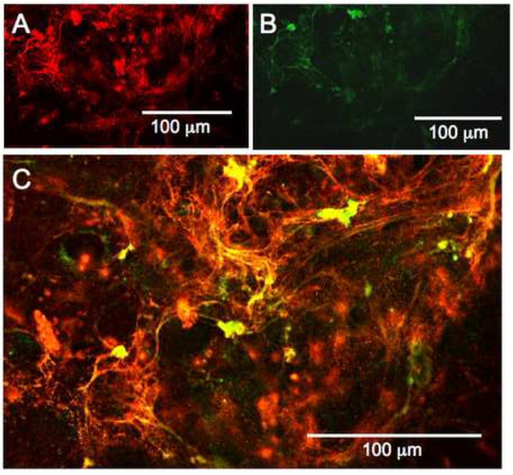
Figure 7. Fluorescently labeled heparin localizes to elastic fibers in vitro.
hAoSMC cultured for 7 days with 300 µg/ml DTAF-heparin. Confocal sections were taken, and different wavelength images of the same z-sections were reported. (A) shows the distribution of labeled heparin. As shown in (B), the DTAF-heparin retains biological activity, as it promotes elastic fiber formation. As seen in (C), the heparin and nascent elastin fibers colocalize extensively.

