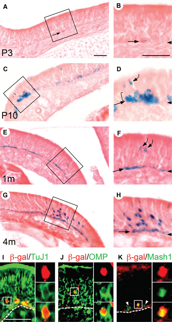
Figure 2.
Horizontal basal cells (HBCs) give rise to neuronal cells in the olfactory epithelium (OE). (A–H): K5.CrePR1 tg/+: R26R+/− mice at P3 (A, B), P10 (C, D)1m (E, F), and 4m (G, H) were examined for β-gal activity by X-gal staining. Insets in (A, C, E, G) are enlarged in (B, D, F, H), respectively. Arrowheads in (B, D, F, H) indicate the position of the basal lamina. (A, B): At P3, infrequent β-gal+ HBCs were observed (arrows). (C, D): By P10, the number of β-gal+ HBCs, as well as the intensity of β-gal activity in HBCs, had increased (straight arrow). Notably, β-gal+ cell clusters containing tentative globose basal cells (GBCs) (bracket) and olfactory receptor neurons (ORNs) (curved arrow) were observed. (E, F): By 1m, the majority of HBCs were β-gal+ (straight arrow). β-Gal+ tentative ORNs were observed (curved arrows). (G, H) At 4m, β-gal+ cell clusters containing many tentative ORNs were observed. (I, J, K): K5.CrePR1 tg/+: R26R+/− mice at 2–3m were examined for β-gal/TuJ1 (I)β-gal/OMP (J), and β-gal/Mash1 (K) expression by double immunofluorescence. Dotted lines indicate the position of the basal lamina. The presence of β-gal+/TuJ1+ (I) and β-gal+/OMP+ cells (J) demonstrated that HBCs had generated ORNs. The presence of β-gal+/Mash1+ cells (K) demonstrated that HBCs had generated Mash1+ GBCs. Arrowheads point to Mash1+ GBCs that are negative for β-gal (K). The red signal in the apical region of the OE corresponds to nonspecific binding of the secondary antibody (K). Scale bars = 60 µm (A, C, E, G)60 µm (B, D, F, H), and 55 µm (I, J, K). Abbreviations: β-gal, β-galactosidase; m, months; OMP, olfactory marker protein; P, postnatal day; TuJ1, neuronal class III β-tubulin.

