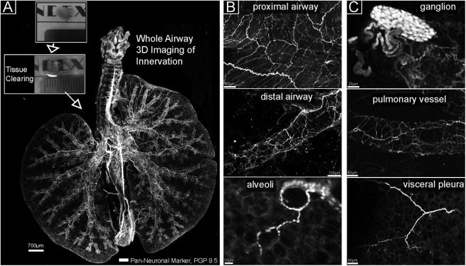
Figure 1.
Tissue clearing and whole airway imaging of nerves. (A) Flattened image of a whole mouse lung. Visible are all nerves stained with the pan-neuronal marker PGP 9.5 (white). The airway is oriented with the left lung on the left and superior/inferior right lung lobes on the right. The boxed areas show a murine lung lobe before (top) and after (bottom) optical clearing. (B, C) Magnified images from A show nerve populations in areas of the lung and a ganglion in the carina region.

