Figure 4.
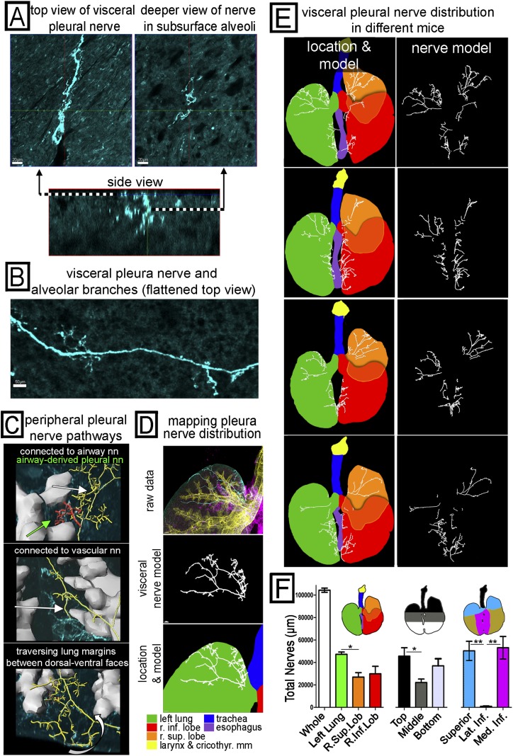
Mapping the pathways of peripheral, visceral pleural nerves and quantifying their overall distribution. (A) A 3D image sample of a visceral pleura nerve is shown in a top-down view of the pleural surface (upper left), a top-down view deeper in alveoli (upper right), and a side view of the same nerve (lower) oriented with the pleural surface on top. (B) A wider field of the pleural surface is shown in a flattened projection to demonstrate a visceral pleural nerve with branches (these usually penetrate alveoli). (C) Some of the novel peripheral pathways taken by visceral nerves are shown with airways (gray) and visceral nerves that have been identified and modeled (yellow, red). Visceral nerves are seen connecting to airway innervation (top, white arrow), connecting to vascular innervation (middle, white arrow), and traversing the lung margins (bottom, white arrow) connecting dorsal and ventral visceral nerve networks. We also found rare patches of visceral nerves (top, green arrow) derived from airway innervation but not connected to the main visceral nerve network. (D–F) Identification, modeling, and mapping of visceral nerve distribution across the whole airway. (D) Views of the superior left lung visceral pleural nerves in cyan from Figure 3A (top), the identified visceral pleural nerves (middle), and visceral pleural nerves overlaid on a lung diagram color coded by region (bottom). The color legend is shown. (E) Visceral pleural nerves shown with (left) and without (right) the color-coded lung diagram showing different regions in four different mouse lungs. (F) Aggregate nerve length based on three different schemes to subdivide the airways (lobe, top to bottom, and superior versus medial inferior versus lateral inferior) was quantified by the computer (n = 4). *P < 0.05; **P < 0.01. Visceral nerve pathways are shown in Videos E4–E6.

