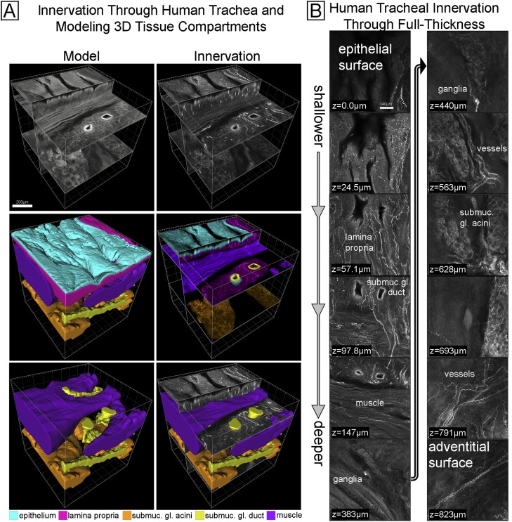
Figure 6.
Imaging innervation and modeling tissue compartments through full-thickness human trachea. (A) A full-thickness 3D image of innervation (upper right) and colored modeled tissue compartments (lower four images) made from red emission autofluorescence (upper left). The middle left and lower left images show the computer model of each tissue compartment. The right middle image shows the nerve image separated (by color) into distinct tissue compartments using the computer model. A color legend for the tissue compartments is included. (B) Select images of the data from A are shown from epithelium (top left) to the adventitia (bottom right). The presence of innervation in tissue compartments is labeled (e.g., lamina propria). See Video E7.

