Figure 5.
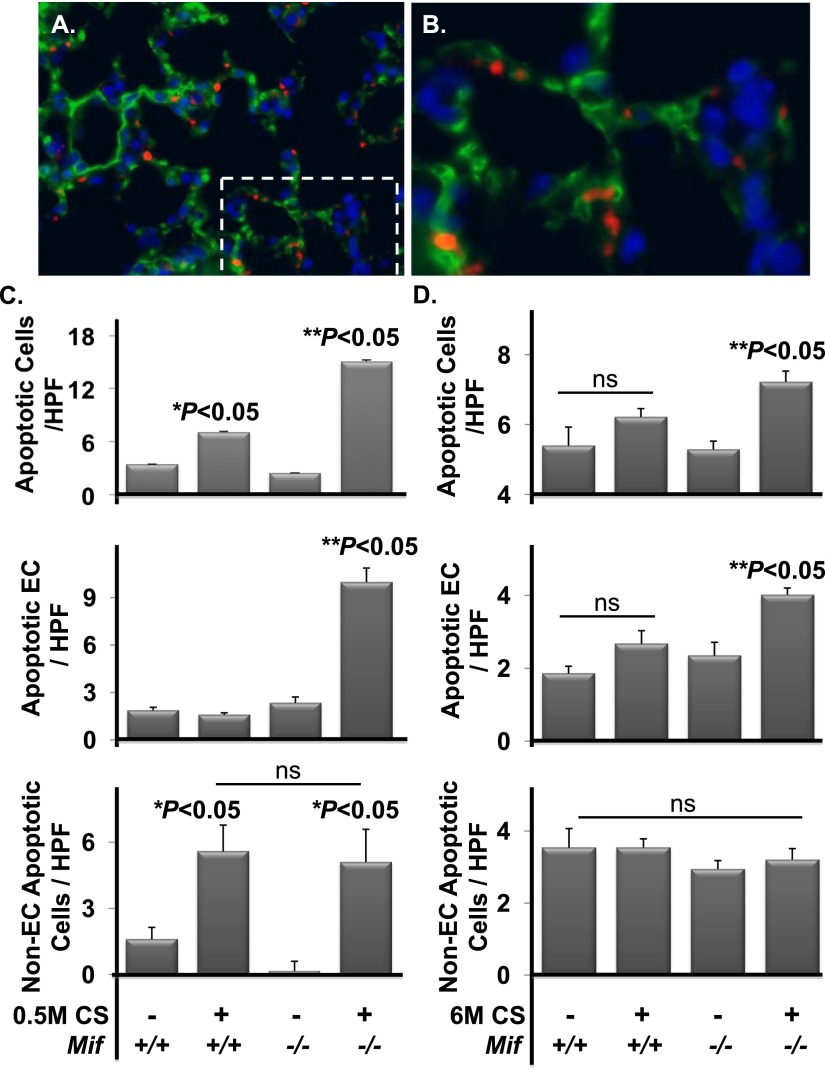
Caspase-3 expression is increased in the absence of MIF with subacute and chronic CS exposure. Lungs from Mif+/+ and Mif−/− animals exposed to filtered air or CS for 0.5 or 6 months were harvested and sectioned for immunohistochemistry. Representative fluorescent microscopy images are shown of cleaved caspase-3 (red) and thrombomodulin (green) in the lung (A and B). Area of magnification denoted with dashed lines (A). The frequency of cleaved caspase-3–positive parenchymal cells was significantly increased in Mif−/− versus Mif+/+ mice exposed to 0.5 months of CS (P < 0.05) ([C] upper panel), and with 6 months of CS (P < 0.05) ([D] upper panel). The majority of caspase-3–positive cells in Mif−/−exposed to 0.5 or 6 months of CS were ECs (both with **P < 0.05) (middle panels [C and D]). Non-ECs were enhanced with 0.5 months with exposure (*P < 0.05), and this did not differ with genoptype (lower panels [C and D]). No differences were observed under basal conditions independent of genotype (n = 5–8 per arm). Nuclei were stained with 4′,6-diamidino-2-phenylindole (blue). Values are expressed as means ± SEM. ns, not significant. *P < 0.05 air versus CS. **P < 0.05 Mif+/+ versus Mif−/−.

