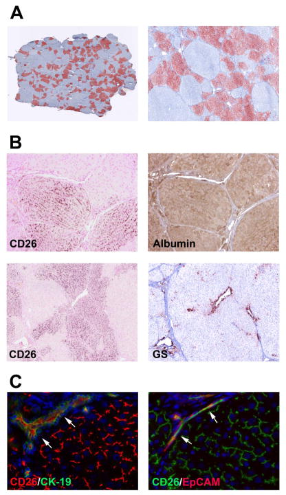
Figure 7.
Repopulation and functional incorporation of differentiated, donor-derived cells after transplantation without PH. ED15 fetal liver cells (~1.8 × 10⁸ cells) were transplanted into DPPIV− rats 3 months after starting TAA administration (A). Images contain 65 or 25 tiled microscopic fields (original magnification, x50 and x100). (B) Patterns of DPPIV (CD26) and albumin expression, and DPPIV and glutamine synthetase (GS) expression in consecutive liver sections demonstrate that DPPIV+ cells differentiated into hepatocytes and become incorporated into morphologically normal hepatocyte plates. (C) Transplanted FLSPCs showed a canalicular pattern of CD26 staining characteristic of mature hepatocytes and also differentiated into bile ducts coexpressing CD26 with CK-19 or EpCAM (see arrows). Original magnification, x10 and x5 (B), x40 (C).

