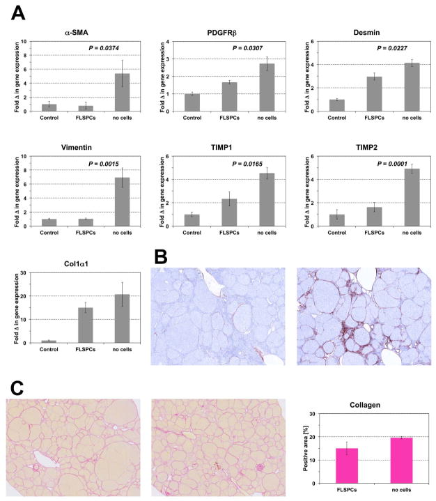
Figure 8.
Effect of FLSPC transplantation on hepatic fibrogenesis. ED15 fetal liver cells (~1.8 × 10⁸ cells) were transplanted into DPPIV− rats 3 months after starting TAA administration, which was continued for 2 months thereafter. Five weeks later, rats were sacrificed. (A) Quantitative RT-PCR analysis for mRNA of genes for stellate cell activation and fibrogenesis. Values are mean ± SEM of liver samples from FLSPC-transplanted fibrotic rats (n=6) or non-cell transplanted fibrotic rats (n=6) compared to age-matched normal control rats, set at a value of 1 (n=3). One representative example from at least 3 replicate experiments is shown. (B) Immunohistochemical detection of α-SMA-positive cells in rat livers with (left) or without (right) cell transplantation. Images contain 25 adjacent microscopic fields (original magnification, x100). (C) Selected areas of Sirius Red-Stained tissue sections of rat livers with (left) or without (middle) cell transplantation. Quantification of Sirius Red-Stained collagen (right). Values are mean ± SEM of whole liver sections (n=6/6).

