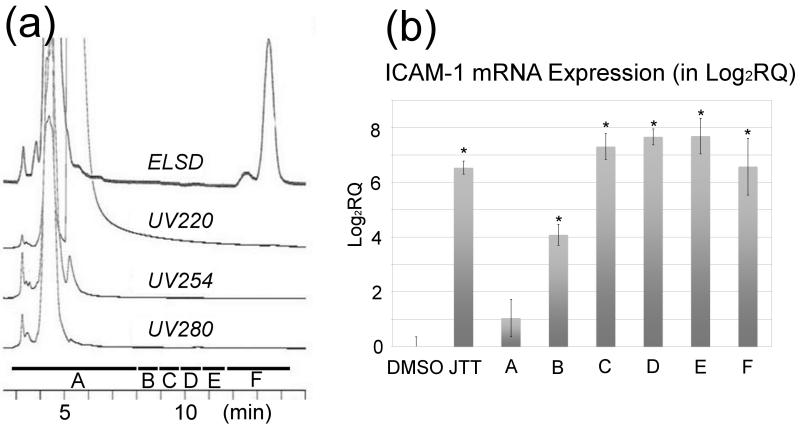
Fig. 2.
Analysis of the fraction (“MeOH” fraction) enriched with the MΦ-stimulatory activity. (a) HPLC (C18) chromatogram of the “MeOH” fraction obtained from the solid phase extraction (C8) in Fig. 1. The HPLC eluent was monitored by PDA detector (at 220, 254, 280 nm) as well as by ELSD. (b) qRT-PCR analysis of THP-1 cells treated with the HPLC fractions. DMSO (vehicle control), JTT (100 μg/mL), LPS (Lipopolysaccharides, positive control, 0.5 μg/mL), all fractions (5 μg/mL). The maximum MΦ-stimulatory activity was eluted between 9 and 12 minutes (Fractions C, D, and E) before the ELSD visible peaks. At least triplicate experiments (n=3) were carried out for each sample. **P < 0.001, compared with the DMSO control.

