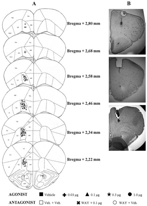
Fig. 1.
a Schematic representation of successive coronal sections of the mouse brain showing the histological verification of injection placement into the VO PFC (n=63) (rostral to caudal: 2.80, 2.68, 2.58, 2.46, 2.34, and 2.22 mm anterior to the bregma). VO ventral orbital cortex; Cgl cingulate cortex, area 1; PrL prelimbic cortex; MO medial orbital cortex; LO lateral orbital cortex; M1 primary motor cortex; M2 secondary motor cortex. All images are adapted from Paxinos and Franklin (2001). b Photomicrographs showing correct placement of guide cannula and microinjection at VO PFC region

