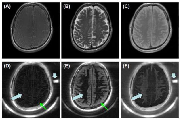
Figure 5.
T1-FSE (A), T2-FSE (B), PD-FSE (C) as well as IR-dUTE imaging of the ultrashort T2 components in white matter of the brain of a healthy volunteer (D–F). Dual echo images were acquired with TEs of 8 μs (D) and 4.4 ms (E). The later echo image was subtracted from the first one to selectively depict short T2* components in white matter (F). Signals from the long T2 component in white matter were nulled by the adiabatic inversion pulse (thick arrows). Signals from the long T2 gray matter were suppressed through subtraction (thin arrows), leaving signal from tightly bound myelin water to be selectively depicted (F). A low RPD of 3.17 ± 0.28% was demonstrated for the ultrashort T2* components by comparing their signal intensities with that of the rubber phantom (short thick arrows).

