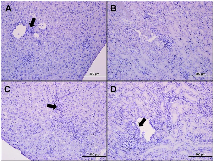
Figure 6. Histopathology of the liver and kidney following lethal intravenous Orientia challenge at days 9 and 12 pi.
Portal triaditis (A-arrow; 20×) was prominent at 9 dpi, and at 12 dpi perivascular infiltrates were observed in the hepatic sinusoids (B-arrow; 20×). Mild perivascular infiltrates were observed in the kidney at 9 dpi (C-20×), and at 12 dpi (D-arrow; 20×) cellular infiltrates were observed throughout the kidney, particularly as peritubular infiltrates.

