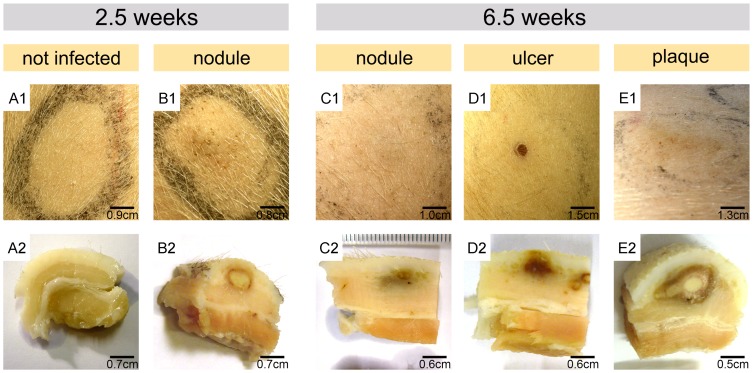
Figure 1. Macroscopic appearance of pig skin infected with M. ulcerans.
Development of representative lesions 2.5 weeks or 6.5 weeks after subcutaneous infection with 2×10⁷ (B1, D1 and E1) or 2×10⁶ (C1) CFU is depicted and compared to a site left uninfected (A1). Excised tissue specimens were fixed and vertically cut in half to visualize macroscopically visible alteration in tissue structure (A2–E2). For high inoculation doses (≥2×10⁶ CFU) the formation of nodules (B1) with necrotic centres was observed already 2.5 weeks after infection (B2). These yellow centres indicative for coagulative necrosis were surrounded by a reddish ring (B2). At 6.5 weeks after infection, these nodules had progressed into a small ulcer or a plaque (D1, E1) associated with marked macroscopically visible alterations in tissue structure (D2, E2). At sites injected with 2×10⁶ CFU nodules with greyish discoloration of the dermis had developed 6.5 weeks after injection of M. ulcerans (C1, C2).

