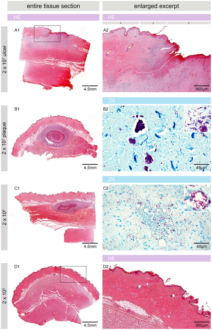
Figure 3. Microscopic appearance of pig skin 6.5 weeks after experimental infection.
Histologic sections stained with Haematoxylin/Eosin (HE) (A1, A2, B1, C1, D1 and D2) or Ziehl-Neelsen/Methylene blue (ZN) (B2, C2). At sites injected with 2×10⁷ CFU nodules had either developed into a small ulcer with destroyed epidermis (z), strong infiltration and indications for the development of undermined edges (A1, A2, dotted line) or into a plaque with a necrotic core surrounded by infiltrating cells (B1). x: intact epidermis, y: epidermal hyperplasia, z: destroyed/missing epidermis. The site infected with 2×10⁶ CFU showed a similar architecture as the plaque but flatter, less organized and with a smaller overall circumference (C1). Both lesions comprised AFB in their necrotic cores, either in big clumps (B2) or in smaller numbers and smaller aggregations (C2). No signs of infection, inflammation and pathology were observed at sites inoculated with 2×10⁵ CFU (D1, D2) or less (not shown).

