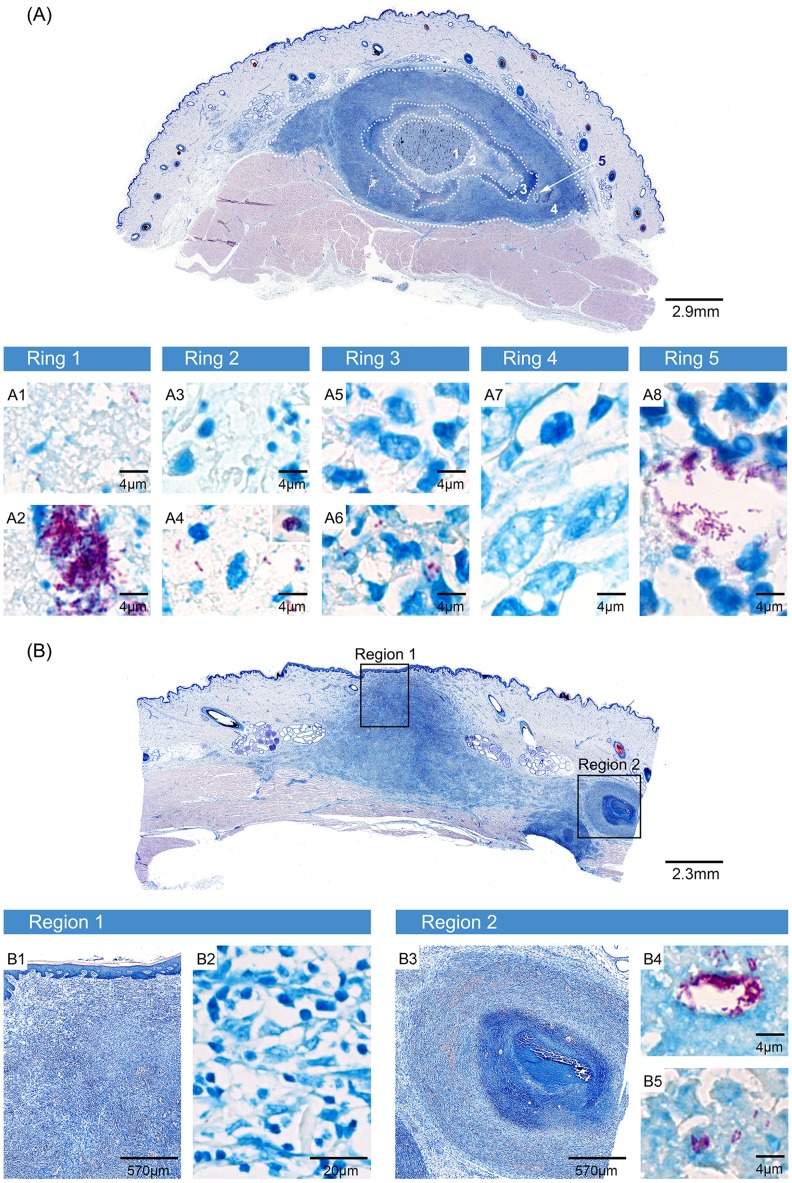
Figure 4. Containment of large amounts of AFB in the necrotic core and development of satellite microcolonies.
Histologic sections stained with ZN. A plaque (A) and a small ulcer (B) are shown that developed 6.5 weeks after infection with 2×10⁷ CFU. The ulcerated lesion was strongly infiltrated at the site of ulceration, where no AFB were found (Region 1, B1, B2). Lateral and between dermis and muscle tissue infiltrating cells enclosed small necrotic areas (Region 2, B3), where AFB were found as satellite microcolonies (B4, B5). The plaque consisted of distinct layers of infiltrating cells encasing a necrotic core containing large clumps of bacteria (Ring 1, A1, A2). A second and third ring with decreasing bacterial load and integrity and increasing integrity of infiltrating cells were layered around this core (Ring 2, A3, A4 and Ring 3, A5, A6). A belt of intact cells was surrounding these three inner layers. It did not contain any AFB (Ring 4, A7) except for a microcolony peripheral to the main bacterial burden (Ring 5, A8).

