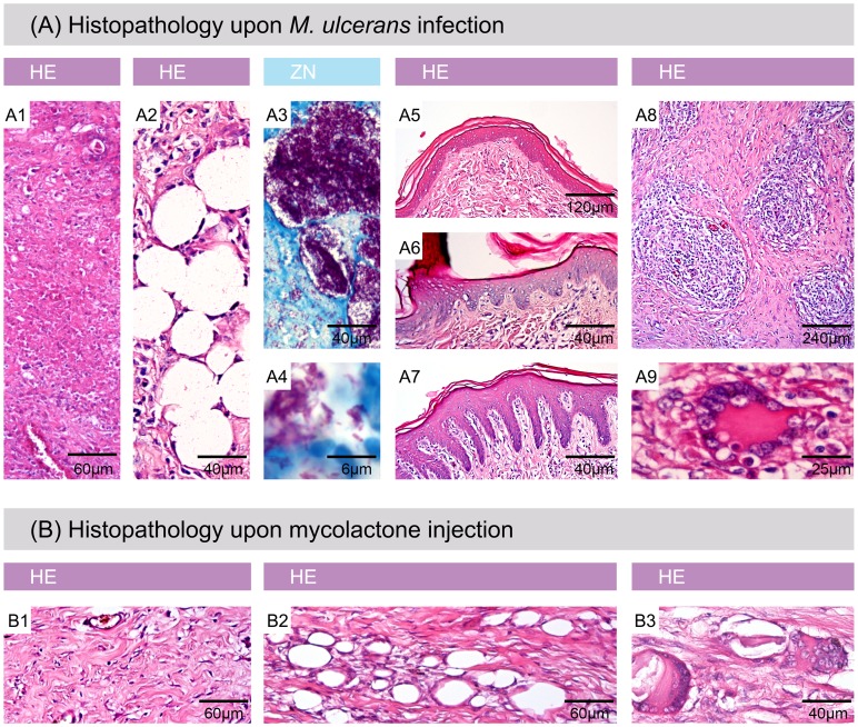
Figure 5. Histophathological hallmarks of Buruli ulcer in experimentally infected pig skin.
Histologic sections stained with Haematoxylin/Eosin (HE) (A1, A2, A5, A6, A7, A8, A9, B1, B2 and B3) or Ziehl-Neelsen/Methylene blue (ZN) (A3, A4). A: All typical histopathological features of BU in humans were found in infected pig skin. A1: necrosis, A2: fat cell ghosts, A3 and A4: extracellular clusters of AFB, A5: healthy epidermis, A6: moderate epidermal hyperplasia, A7: strong epidermal hyperplasia, A8: granuloma formation, A9: giant cells. B: Histopathological changes induced by mycolactone injection. B1: necrosis, B2: fat cell ghosts, B3: giant cells.

