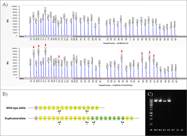
Figure 2.

Results of the molecular analysis on the CHEK2 duplication. (A) Electropherograms showing the MLPA probes for CHEK2 exons in a reference sample (upper panel) and in proband III-4 (lower panel): duplicated exons are indicated by red arrows (Coffalyser software uses the NM_001005735.1 transcript of CHEK2). (B) Schematic representation of CHEK2 gene showing the wild type allele and the duplicated allele (transcript NM_007194.3 with non-coding exon 1 in grey): arrows indicate primers 13-forward and 6-reverse used for PCR. (C) Agarose gel showing the breakpoint of the duplication amplified by PCR: the reaction generated a 6-kb product in patients with the duplication (II-3, II-5, III-4 and III-5) but not in the wild type individual (II-7) or negative controls (C1 and C2). M, molecular weight size marker 1 kb (Promega).
